All products

SmallAir™ (SA)

General features of SmallAir™

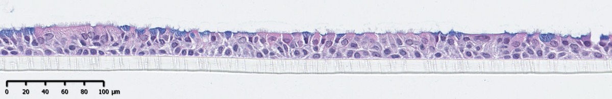
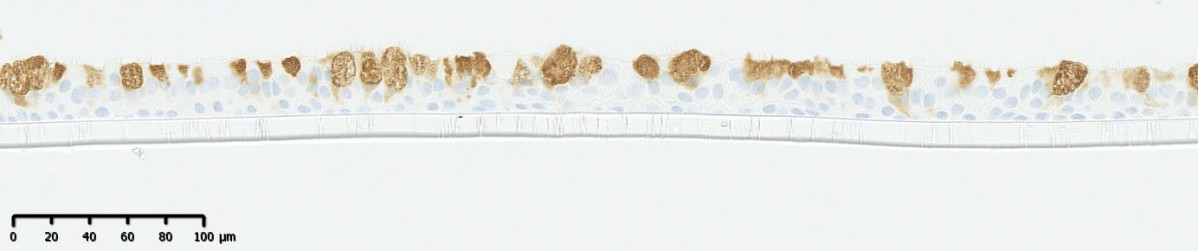
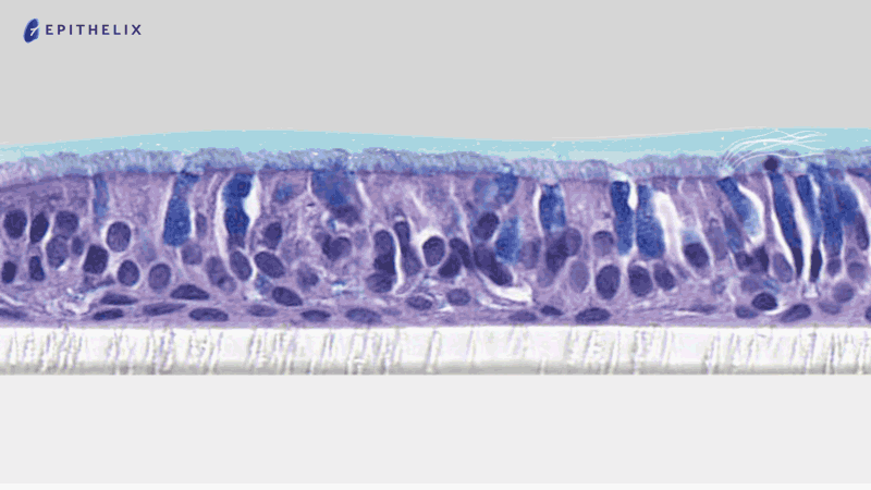
SmallAir™ is an in vitro tissue model of the human lower airway epithelium (bronchiolar region after the 5th division of the airway) cultured at the air liquid interface (ALI). It is a powerful and predictive model for in vitro research and tests. SmallAir™ tissue is mainly composed of the following cell types:

  • Basal cells (progenitor cells)
  • Club cells (cuboidal cell, known to be metabolically active cells)
  • Ciliated cells (with active cilia)
  • Goblet cells (present in low proportion, mucus producing cells)

Figure 1: schematic representation of a SmallAir™ cross section

A meticulous tissue production

Since 2017 we produce SmallAir™ with high standards of quality and reproducibility. The tissue production is GIVIMP certified since 2023.

To ensure high predictability, SmallAir™ is solely reconstituted using low passage (P1), human primary cells. The primary cells are isolated from micro dissection of the bronchioles of human lung samples, under ethical approval and donor consent. Each batch of tissue is delivered with a certificate of Analysis (COA) with donor information and Quality Control (QC) results.

Patients with several pathologies are available for SmallAir production

  • Healthy (non-smoker)
  • Healthy (smoker)
  • COPD (Chronic Obstructive Pulmonary Disease)
  • Asthmatic
  • Allergic Rhinitis

SmallAir™ recapitulates the functions of the human small airway epithelium

Our models mimic the characteristics of in vivo tissues:

Long shelf life

Once the production cycle is terminated, SmallAir™ is in a stable homeostatic state for several month with a slow but physiological cell renewal. It maintains its functionality and cellular population during its entire life cycle.

Basal, Club, Ciliated and Goblet cells are the main cell type constituting SmallAir™. The major difference from the upper airway is the abundant presence of club cells, quasi absent in the upper airways. On the other hands goblet cells are present but in low proportion.

Figure 2: histological cross section of SmallAir™ with Alcian blue H/E staining of the tissue allowing to visualize the tissue structure

Club cells:

Figure 3: histological cross section of SmallAir™ with CC10 staining of the tissue allowing to visualize the club cells.

Basal cells:

Figure 4: CC-10 was detected by Western Blot. As expected, SmallAir™ contained few Muc5-Ac positive cells (goblet cells). In contrast, CC-10 was not detected in MucilAir™. Instead, Muc-5Ac was highly expressed in MucilAir™.

Functional cilia

As observed on the human tissue a dense cilia layer is present on the surface of SmallAir™.

The Cilia Beating Frenquency (CBF) could be monitored with our tailored software (Cilia-X) and is part of the SmallAir™ QC.

Mucus production

Goblet cells produce and secrete mucus on the apical side of the tissue. The mucus layer is therefore produced by the tissue itself in a physiological, complex and dynamic manner.

Figure 6: Schematic animation of the mucus release on the apical side

Mucociliary clearance

Thanks to the presence of functional cilia beating cells and the mucus layer, a functional mucociliary clearance is present on top of SmallAir.

Figure 7: Mucociliary clearance assay with software tracking of polystyrene for quantitative analysis

Tight junctions

Being the first barrier in the airway the tissue is naturally tight thanks to the presence of tight junctions.

Barrier function could be quantified with a Trans Epithelial Electrical Resistance (TEER) device, and is part of the SmallAir QC.

Native cell surface receptor and ion channels

Using low passage cells allows to keep the native ion channel (CFTR, ENAC, etc.) allowing the use of the model in electrophysiology experiments.

Native cell surface receptors (TMPRSS2, ACE2, FCRn, etc.) are also present. This makes the model an efficient tool to study viral infections.

Release of soluble factors

SmallAir is also a very potent immune-regulator and secrete a wide panel of Cytokines, Chemokines and, Metalloproteinases that could be present at a constitutive level but also in stimulated condition.

An easy and ready-to-use model

We handle all the production and QC process to deliver SmallAir ready-to-use at your lab. No fancy equipment or cell culture skills are required for its maintenance.

Once received, transfer the tissues in fresh medium and in the incubator. Then change the medium twice a week, occasionally perform a mucus wash. That’s it.

SmallAir™ culture medium, a dedicated, chemically defined and serum-free culture medium ensures a stable composition from batch to batch.

Need technical support for your experiment with SmallAir™? Contact us!

Easy shipping

We ship routinely our tissues to Europe, America and Asia. Our packaging method allows to deliver safely the tissues to these locations.

We can evaluate, with you, the delivery to your place.

SmallAir™ is widely referenced in the literature

More than +800 scientific publications using Epithelix tissues.

Check out our publication page!